Leg Muscles Diagram / Zyce59 Ibegtrm - For women, shaping the thigh muscles is an essential goal of physical fitness.. The lower leg lies between the knee and the ankle. The short head originates from the lateral lip of linea aspera and. Most leg pain results from wear and tear, overuse, or injuries in joints or bones or in muscles, ligaments, tendons or other soft tissues. The 3 muscles are called triceps coxae. There are four muscles in this compartment:
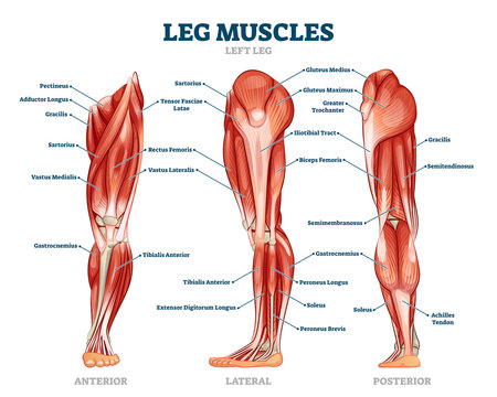
Legs are used for standing, and all forms of. These four muscles at the front of the thigh are the major extensors (help to extend the leg. Spend some time revising this diagram by connecting the name and location of each structure with what you've just learned in the video. Leg muscles labeled take a look at the leg muscles diagram below, where you see each muscle clearly labeled. The muscles that make up the quadriceps are the strongest and leanest of all muscles in the body.
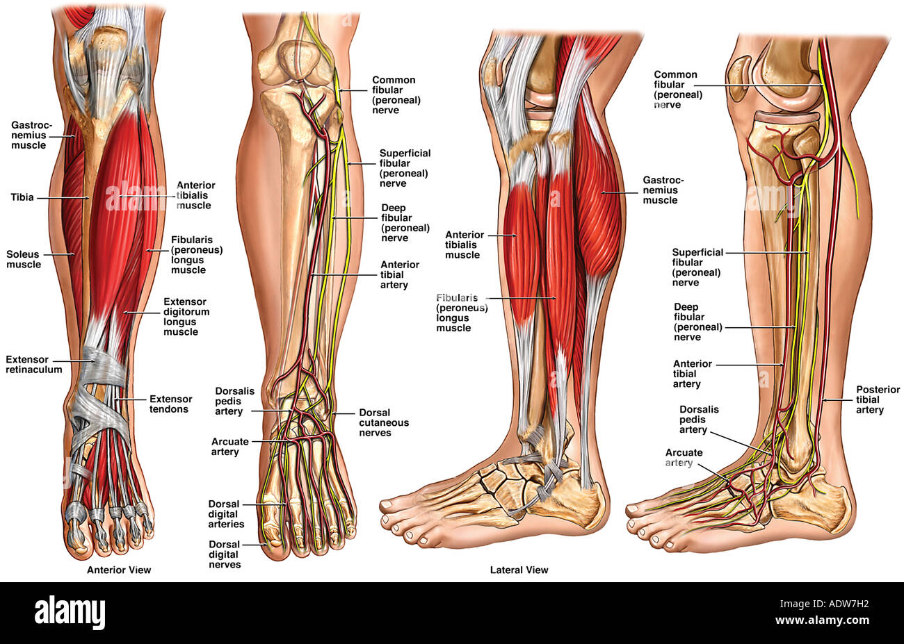
Leg Muscles Diagram Anterior Leg Muscles Gray S Anatomy Illustration Radiology Case Radiopaedia Org Two Heads Extends From The Ischial Tuberosity To The Rendi Wijayanto from i1.wp.com Pain in your calf or thigh can be caused by muscle cramps, a pulled or strained muscle, or issues related to your nerves. Included are several layered views of the back muscles, the dorsal muscles, subclavius muscles, rhomboideus major and minor muscles, deltoid muscles and many more. Flexes elbow and moves forearm. The calf muscle, on the back of the lower leg, is actually made up of two muscles: The long head arises from a common tendon with semitendinosus from the superior medial quadrant of the posterior portion of the ischial tuberosity. These muscles include the gluteus maximus muscle (the largest muscle in the body) and the hamstrings group, which consists of the biceps femoris, semimembranosus, and semitendinosus muscles. Leg pain can also be caused by blood clots, varicose veins or poor circulation. Each of these major nerves further divides into many smaller nerve branches to stimulate individual muscles and sense touch, pain, warmth, and cold in the skin.
One of the most important tendons in terms of mobility of the leg is the achilles tendon.
It acts as a tensor of the arches of the foot, but can also be added with the first digit and plantar flexion of its first phalanx. Anterior compartment thigh muscles this is the largest of the three compartments of the thigh. The following diagram illustrates the actions of the terms adduction, abduction, flexion and extension at the different joints. The largest muscle masses in the leg are present in the thigh and the calf. For images of the muscle, click on each link under location. The muscles in the hip are responsible for the movement of the hip and, by proxy, the leg. The 3 muscles are called triceps coxae. Human anatomy diagram from the back view 12 photos of the human anatomy diagram from the back view human anatomy diagram back view organs, human anatomy diagram rear view, human muscles, human anatomy diagram back view organs, human anatomy diagram rear view. Included are several layered views of the back muscles, the dorsal muscles, subclavius muscles, rhomboideus major and minor muscles, deltoid muscles and many more. The muscles work together to enable movement and keep the hip in alignment. This important tendon in the back of the calf and ankle stores the elastic energy needed for running, jumping, and other physical activity. A muscle located on the back portion of the lower leg, being one of the two major muscles that make up the calf:the flexing of this muscle during walking and bending of the knee creates traction on the femur, pulling it toward the tibia in the lower leg and causing the knee to bend. Notice the upper leg has a biceps muscle just like the upper arm does.
Brings leg back to and across body. Biceps femoris (long head) biceps femoris (short head) semitendinosus. The gastrocnemius is the larger calf muscle, forming the bulge visible beneath the skin. Brings hip away from body. The following diagram illustrates the actions of the terms adduction, abduction, flexion and extension at the different joints.
Lower Leg Anatomy High Resolution Stock Photography And Images Alamy from c8.alamy.com Muscle and bone anatomy 12 photos of the muscle and bone anatomy back muscles and bones anatomy, human muscle and bone anatomy, muscle & bone anatomy 3d free download, muscle and bone anatomy app, muscle and bone anatomy quiz, human muscles, back muscles and bones anatomy, human muscle and bone anatomy, muscle & bone. It acts as a tensor of the arches of the foot, but can also be added with the first digit and plantar flexion of its first phalanx. Notice the upper leg has a biceps muscle just like the upper arm does. For women, shaping the thigh muscles is an essential goal of physical fitness. The following diagram illustrates the actions of the terms adduction, abduction, flexion and extension at the different joints. The muscles that make up the quadriceps are the strongest and leanest of all muscles in the body. These muscles include the gluteus maximus muscle (the largest muscle in the body) and the hamstrings group, which consists of the biceps femoris, semimembranosus, and semitendinosus muscles. Human anatomy diagram from the back view 12 photos of the human anatomy diagram from the back view human anatomy diagram back view organs, human anatomy diagram rear view, human muscles, human anatomy diagram back view organs, human anatomy diagram rear view.
The calf muscle, on the back of the lower leg, is actually made up of two muscles:
Flexes elbow and moves forearm. The calf muscle, on the back of the lower leg, is actually made up of two muscles: The muscles in the hip are responsible for the movement of the hip and, by proxy, the leg. A muscle located on the back portion of the lower leg, being one of the two major muscles that make up the calf:the flexing of this muscle during walking and bending of the knee creates traction on the femur, pulling it toward the tibia in the lower leg and causing the knee to bend. Leg muscles labeled take a look at the leg muscles diagram below, where you see each muscle clearly labeled. The muscles that make up the quadriceps are the strongest and leanest of all muscles in the body. Extends spine and trunk back. The groin muscles are a group of muscles situated high on the leg in the inner thigh. See more ideas about muscle anatomy, human anatomy and physiology, body anatomy. Learn vocabulary, terms, and more with flashcards, games, and other study tools. There are many muscles located in the lower leg, but there are three that are particularly well known—the gastrocnemius and the soleus, which are the most powerful muscles in the lower leg, and the anterior tibialis. This is why you have to indicate which biceps you are taking about when discussing one or other of these muscles. The following diagram illustrates the actions of the terms adduction, abduction, flexion and extension at the different joints.
Collectively referred to as the hip adductors, the groin muscles are responsible for adduction of the hip, or drawing the leg in. This important tendon in the back of the calf and ankle stores the elastic energy needed for running, jumping, and other physical activity. Notice the upper leg has a biceps muscle just like the upper arm does. The calf muscle, on the back of the lower leg, is actually made up of two muscles: Brings hip away from body.
13 845 Best Leg Muscle Anatomy Images Stock Photos Vectors Adobe Stock from t4.ftcdn.net It is also visible on the medial edge of the thigh from the anterior. The muscles in the hip are responsible for the movement of the hip and, by proxy, the leg. They receive their innervation via the deep. Flexes elbow and moves forearm. Some common causes of leg pain include: The biceps femoris is a muscle of the posterior thigh composed of a long head and a short head. Like the quadriceps, the hamstring muscle group also contains four separate muscles: The hip muscles work together to carry out 4 different types of movement:
Observe the leg muscle diagram posted above and notice that there are many parts in the muscles.
Muscle of the frog by label Learn vocabulary, terms, and more with flashcards, games, and other study tools. Your leg muscles are some of the hardest working muscles in your body. The quad muscles— which form the meaty mass on the front of your thighs — are among your strongest muscle groups, and play a critical role in athletic activities. This anatomy chart is a great example of beauty and function in one, as it is pleasing to look… This important tendon in the back of the calf and ankle stores the elastic energy needed for running, jumping, and other physical activity. There are many muscles located in the lower leg, but there are three that are particularly well known—the gastrocnemius and the soleus, which are the most powerful muscles in the lower leg, and the anterior tibialis. Human anatomy diagram from the back view 12 photos of the human anatomy diagram from the back view human anatomy diagram back view organs, human anatomy diagram rear view, human muscles, human anatomy diagram back view organs, human anatomy diagram rear view. The hamstring muscles, also known as the rear thighs, make up the backside of the upper leg anatomy. Brings leg back to and across body. Legs are used for standing, and all forms of. These four muscles at the front of the thigh are the major extensors (help to extend the leg. The following diagram illustrates the actions of the terms adduction, abduction, flexion and extension at the different joints.